-
Histology Photomicrographs
Human Anatomy and Physiology (BIOL& 241L-242L)
Karen Hart, Peninsula College
Cardiac muscle

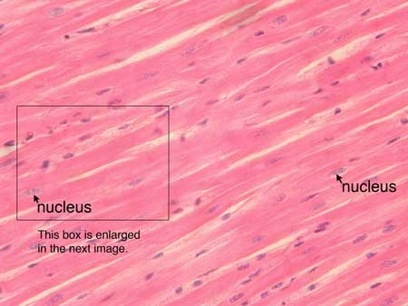
Slide: cardiac muscle HD 4-22
Microscope at 400X
Cardiac muscle cells are cylindrical cells whose ends branch and form junctions with other cardiac muscle cells. A cardiac muscle cell typically has one nucleus located near the center. Two cardiac muscle cell nuclei are indicated in the labelled image. Many more cardiac muscle cell nuclei are visible. In the connective tissue between cardiac muscle cells are nuclei of other cells such as fibroblasts and endothelial cells of capillaries.
Cardiac muscle cells contain myofibrils whose sarcomeres are more or less aligned, however the striations of a cardiac muscle cell are much fainter than the striations visible in a skeletal muscle cell. In the images of cardiac muscle on this page striations are not visible.
Cardiac muscle cells are joined to each other by junctions called intercalated disks. Arrows in the enlarged image indicate several intercalated disks. Each intercalated disk appears as a darker staining line across the cell. An intercalated disk contains desmosomes which mechanically hold the cells to each other and also contains gap juctions which allow ions to pass freely between cells. The presence of gap junctions means that an action potential in one cardiac muscle cell will cause of flow of ions (a local current) through gap junctions into adjacent cells. The local current will set off an action potential in the adjacent cells. Thus an action potential in one cardiac muscle cell quickly spreads to other cardiac muscle cells.
Slide: cardiac muscle HD 4-22
Microscope at 400X

This image is an enlargement of the box in previous image.
Unless otherwise noted, contents © 2006-2026 Karen Hart