-
Histology Photomicrographs
Human Anatomy and Physiology (BIOL& 241L-242L)
Karen Hart, Peninsula College
Cartilage and bone: Spongy bone
Spongy bone is the tissue that makes up the interior of bones; compact bone is the tissue that forms the surface of bones. In long bones, spongy bone forms the interior of the epiphyses; the diaphysis (shaft) consists of compact bone surrounding the central marrow cavity.
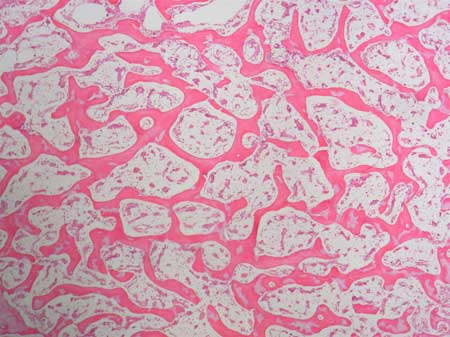
Slide: Cancellous bone HB 8-311
Microscope at 40X
Spongy bone is a network of irregularly-shaped sheets and spikes of bone (trabeculae). The trabeculae are only a few cell layers thick. The spaces between the trabeculae contain red or yellow marrow, depending on a person's age and on which bone it is.
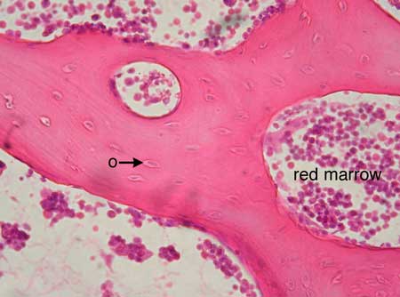
The marrow in these images is red marrow. Red marrow contains blood stem cells and blood cells in all stages of development. These cells are supported by a reticular connective tissue framework. In yellow marrow one would see abundant adipocytes instead of
blood-forming cells.
The arrow labeled o points to an osteocyte in its lacuna. Canaliculi (not visible here) connect the lacunae of osteocytes to each other and to the marrow spaces between the trabeculae.
There are no blood vessels within the matrix of spongy bone, but blood vessels are nearby in the marrow spaces. Exchange of nutrients, gases, etc. occurs between capillaries in the marrow and the interstitial fluid of the marrow. The interstitial fluid extends into the canaliculi and thereby supplies the osteocytes.

Slide: Cancellous bone HB 8-311
Microscope at 400X
Slide: Cancellous bone HB 8-311
Microscope at 400X
Unless otherwise noted, contents © 2006-2026 Karen Hart