-
Histology Photomicrographs
Human Anatomy and Physiology (BIOL& 241L-242L)
Karen Hart, Peninsula College
Skeletal muscle tissue
The muscles that attach to the skeleton are called skeletal muscles. Contraction of skeletal muscles maintains posture, produces movement of the whole body (walking, running, etc.) and produces movements of parts of the body (moving the hand, moving the eyes, smiling, etc.). Contraction of skeletal muscles is under voluntarily control.

Slide: Hair follicle sec. 3 types HI 1-3
Microscope at 400X

The top and middle images show skeletal muscle tissue in longitudinal section. Skeletal muscle cells (also known as skeletal muscle fibers) are cylindrical, multinucleate and very long. The images show only part of length of these skeletal muscle cells.
Note the striations (stripes) that run across each cell. This pattern of light and dark is due to the arrangement of the protein filaments involved in muscle contraction.
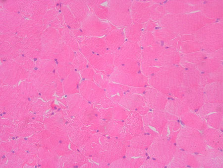
The bottom image shows a cross section of skeletal muscle tissue. Individual skeletal muscle cells appear as round or polygonal shapes separated from each other by a thin layer of connective tissue (the endomysium) which appears white in this slide.
The interior of a skeletal muscle cell is filled with the structures involved in contraction (myofibrils, sarcoplasmic reticulum, T tubules, mitochondria, etc.). The nuclei are pushed out to the periphery of the cell and typically appear as little bulges at the surface of the cell; bulging nuclei (stained purple) can be seen in the top image (a longitudinal section). The bottom image (a cross section) shows that the nuclei (purple) are always at the edges of the cell.
Slide: Striated muscle l.s. HD 2-21
Microscope at 400X
Slide: Skeletal muscle c.s. H6032
Microscope at 400X
Unless otherwise noted, contents © 2006-2026 Karen Hart