-
Histology Photomicrographs
Human Anatomy and Physiology (BIOL& 241L-242L)
Karen Hart, Peninsula College
Smooth muscle tissue
Smooth muscle contraction is not under voluntary control.
Smooth muscle is found, for example, in the:
walls of blood vessels
walls of air passages
walls of hollow organs such as the stomach, intestine, bladder, and uterus
walls of tubes of the reproductive tracts
arrector pili muscles of the skin
iris and ciliary body of the eye
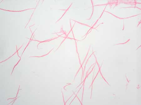
Slide: Smooth muscle (visceral): macerated HD 1-1
Microscope at 400X
The top image shows smooth muscle tissue that has been treated so that the smooth muscle cells separate from each other. An individual smooth muscle cell is very thin and tapers to its ends. Though not visible in this image, each cell has an oval nucleus in the center.
The lower image shows smooth muscle layers of the intestinal wall. In the inner layer, the smooth muscle cells are cut in longitudinal section. The oval nuclei are stained purple. In the outer layer, the smooth muscle cells are cut in cross section; in some cells the cut passes through the nucleus, in other cells the cut does not pass through the nucleus.

Slide: Epithelia 3 Types HA 6-1
Cross section of intestine
Microscope at 400X
Unless otherwise noted, contents © 2006-2026 Karen Hart