-
Histology Photomicrographs
Human Anatomy and Physiology (BIOL& 241L-242L)
Karen Hart, Peninsula College
Kidney cortex

Slide: Epithelia 3 types HA 6-1
Location: Kidney cortex
Microscope at 400X
The kidney cortex consists mainly of closely packed nephrons and blood vessels. A human kidney contains approximately a million nephrons. Each nephron consists of glomerular capillaries, glomerular capsule and renal tubule.
The basement membrane of the glomerular capillaries is fused to the basement membrane of a layer of specialized epithelial cells called podocytes.
The layer of podocytes forms the inner layer of a structure called the glomerular capsule. The outer layer of the glomerular capsule consists of a simple squamous epithelium.
In the first stage of urine formation blood pressure within the glomerular capillaries forces some fluid out of the capillaries, through basement membrane material, through tiny slits between the podocytes and into the glomerular capsule.
This fluid (now called glomerular filtrate) is then moved along the renal tubule of the nephron to be emptied into a collecting duct. As the fluid is moved along, needed substances are reabsorbed and thereby retained in the body. Other substances remain in the tubule lumen and will leave the body in the urine. In addition, certain substances are secreted into the tubule lumen to leave the body in the urine.
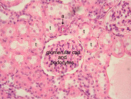
The labeled image above shows closely packed renal tubules (several are labeled t) and a section through a glomerular capsule. A cross section through an arteriole is labeled a.
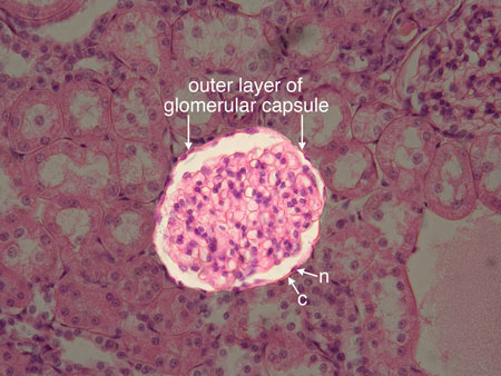
The labeled image above shows the simple squamous epithelium that forms the outer layer of the glomerular capsule. The nucleus of one of the simple squamous epithelial cells is labeled n, the cytoplasm is labeled c.
Unless otherwise noted, contents © 2006-2026 Karen Hart