-
Histology Photomicrographs
Human Anatomy and Physiology (BIOL& 241L-242L)
Karen Hart, Peninsula College
Epithelial tissue: Simple cuboidal epithelium
Example: A simple cuboidal epithelium forms the walls of certain kidney tubules.
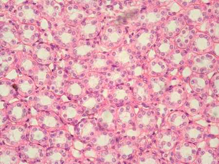
Slide: Epithelia 3 types HA 6-1
Location: Kidney medulla
Microscope at 400X
The upper photo shows a cross section of kidney tubules. The lower photo shows a longitudinal section of kidney tubules.
The wall of a tubule is composed of a single layer of cuboidal cells (a simple cuboidal epithelium).
In the cross section each tubule appears as a ring of cuboidal cells around a white space (the lumen of the tubule).
In the longitudinal section the lumen of a tubule is indicated by lu. Arrows indicate the edges of this tubule. The darker line of pink at the edges is the basement membrane material of the tubule.

Slide: Epithelia 3 types HA 6-1
Location: Kidney medulla
Microscope at 400X
Unless otherwise noted, contents © 2006-2026 Karen Hart